


 Physician & ExpertiseOp. Dr. Cennet Ay TokObstetrics & Gynecology Specialist
 Ultrasound & Imaging · Ordu
The purpose of ultrasound at different pregnancy stages
Ultrasound provides measurements as well as images of the baby. The uterus, placenta and fluid around the baby can also be assessed. Not every structure can be examined in the same detail at every visit: the clinical question changes as pregnancy progresses.
Share your last menstrual period, any earlier reports and how the pregnancy was conceived. Dating information is particularly useful with irregular cycles or assisted conception. Pregnancy follow-up combines ultrasound findings with the wider clinical assessment and relevant laboratory tests.
Early pregnancy: location, number and dating
An early scan may assess the location of the pregnancy, developmental findings and the number of babies. If it is performed very early, expected structures may not yet be visible. That finding alone does not establish a pregnancy problem; a repeat assessment may be needed at an appropriate interval.
Anatomical assessment and growth scans
A detailed mid-pregnancy assessment examines the baby’s anatomy. Later scans may focus on growth measurements, amniotic fluid and the placenta. The timing and need for each examination depend on the initial care plan and any findings that arise.
Ultrasound, screening and diagnostic tests
Ultrasound can identify many structural findings but cannot exclude every condition or genetic disorder. A normal-looking examination does not mean that every problem that could develop later has been ruled out. The baby’s position, gestational age and imaging conditions can affect what is visible.
Measurements such as nuchal translucency may form part of a screening programme. The differing roles of scans and blood tests are explained on the prenatal screening tests page. If a finding needs clarification, the clinician can explain its significance and whether further imaging or specialist assessment is appropriate.
A screening result indicating increased risk is not a diagnosis. If a diagnostic test is proposed, discuss the question it is intended to answer and its limitations. Asking why a test is recommended is a normal part of making an informed decision.
Preparing for the examination
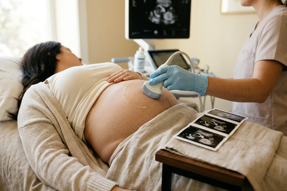
For an abdominal examination, gel is applied to the skin and a probe is moved over the abdomen. In some early pregnancies, an internal scan may be offered for clearer views; the method is explained and your consent is sought. Tell the clinician if you feel uncomfortable during the examination.
Ask whether you need a full bladder, as preparation is not the same at every stage. Bring earlier scan and screening reports. Concentrated silence while measurements are taken does not itself indicate a problem; ask for the findings to be explained when the assessment is complete.
When might a repeat scan or Doppler be considered?
If the baby’s position prevents adequate views, the examination may need to be completed at another appointment. Being called back does not necessarily mean that an abnormality has been found. It is useful to distinguish completed assessments from structures that could not yet be assessed.
Doppler ultrasound may add information when there is a question about growth or placental function. It is not a fixed requirement at every visit for every pregnancy. Follow-up intervals reflect the overall situation and comparison with earlier measurements.
Planning prenatal scan visits in Fatsa, Ordu
When arranging a visit in Fatsa, state your gestational age and why the examination has been requested. If you have been referred for a detailed anatomical assessment, share the referral or previous report. This helps distinguish a routine review from an examination intended to answer a particular question.
If travelling from Ünye, Perşembe or neighbouring inland districts, note the recommended date range should a repeat scan be needed. Keeping reports together supports comparison across different facilities. Before leaving, clarify the next review date and which symptoms should prompt an earlier assessment.
Bring a short list of questions: is the assessment complete, is another test needed and do the findings change the follow-up plan? Ask about unfamiliar abbreviations or measurements and retain the report for subsequent appointments.
Conclusion
Prenatal ultrasound is most useful when it addresses the right question at the appropriate stage. Images and measurements are interpreted alongside gestational age and previous findings. Understanding the scope, limitations and next step helps you prepare for your pregnancy appointments in Ordu.
Clinic Address
The consultation practice is in Fatsa, Ordu. Use the contact details for appointment, location and follow-up information.
Frequently asked questions
Is every prenatal scan a detailed anatomy scan?
No. Routine reviews, early pregnancy scans and detailed anatomical examinations have different scopes. Ask which assessment is planned for your appointment.
Does a 4D image provide a more definite diagnosis?
4D is an imaging format and does not replace a detailed medical assessment. Its usefulness depends on the clinical question and the views that can be obtained.
Do I need to fast before the scan?
Fasting is generally unnecessary for a standard pregnancy scan. Confirm preparation if another test is scheduled that day or you have received specific instructions.
If the scan is normal, do I still need pregnancy appointments?
Yes. Ultrasound is one part of pregnancy care; blood pressure, symptoms and relevant tests also matter. A normal scan does not replace the planned follow-up.
What does it mean if a heartbeat is not seen at the first scan?
A single examination may be inconclusive very early in pregnancy or when dates are uncertain. Measurements, symptoms and a repeat scan when appropriate guide the interpretation.
About this article
Consultation and individual assessment
Op. Dr. Cennet Ay TokObstetrics & Gynecology Specialist · Fatsa, Ordu
Editorial details
- Content preparation
- Editorial information prepared using the medical sources listed below.
- Medical review
- The content update date does not indicate that a physician has medically reviewed this article.
Sources
Medical references used to prepare this article. Links open the original publications in a new tab.
- NHS — Ultrasound scans in pregnancynhs.uk
- ISUOG — Ultrasound Scan in Pregnancyisuog.org
- NICE — Antenatal care NG201nice.org.uk
This article provides general information and does not replace a medical consultation. Diagnosis, treatment suitability and follow-up are determined individually after a clinical assessment.